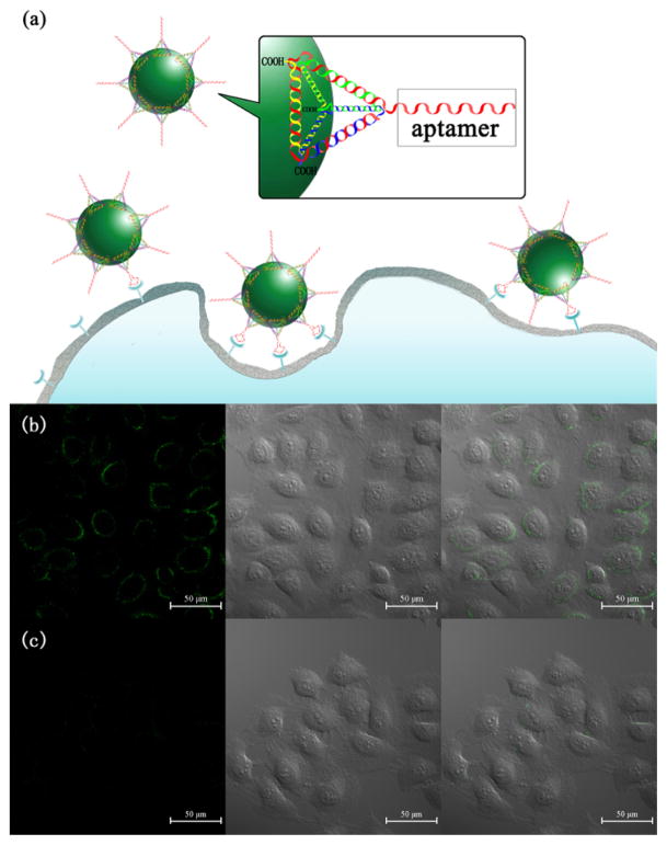
Figure 5.
(a) Targeted imaging of cancer cells with DNA tetrahedron nanostructure-functionalized UCNPs. Confocal microscopy images of MCF-7 cells treated with (b) Apt-tet-UCNPs and (c) Rdm-tet-UCNPs. Each series can be classified as the upconversion luminescent images (left), bright-field (middle), and overlay of both (right), respectively.

