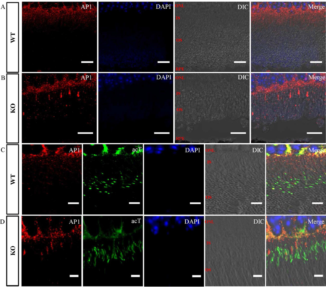
Fig. 7. Mislocalization of the AP1 in Lztfl1 knockout mouse retina.
Immunofluorescent staining of photoreceptor cells in retinas from wild-type (A and C) and Lztfl1 knockout (B and D) mice with antibodies against AP1 (red, A–D) and acetylated tubulin (acT, green, C and D). AP1 was present in the inner segment of all wild-type photoreceptor cells (A). However, it was found to be highly enriched only in some of the photoreceptor cells in Lztfl1−/− mice (B). Acetylated tubulin and AP1 co-staining showed that the abnormally enriched AP1 in knockout photoreceptor cells was partially associated with the major connecting cilium structure (D). WT, wild-type; KO, knockout.

