
Figure 4.
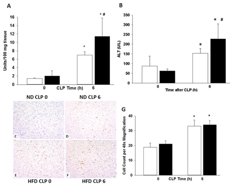
Mice were randomized to a HFD or ND for 6–7wks. Polymicrobial sepsis was induced by CLP after diet intervention and (A) liver neutrophil infiltration was determined by myeloperoxidase assay. (B) Plasma ALT levels were measured. Representative immunohistochemistry for CD68 of liver sections from mice on (C) ND prior to CLP (D) on ND at 6h after CLP (E) on HFD prior to CLP and (F) on HFD at 6h after CLP. Images at 20x magnification and (G) quantification performed at 40x. *p<0.05 vs. time 0h. #p<0.05 vs. normal diet. White boxes = Normal diet, Black boxes = HFD. n=5–8 mice/group.