
Figure 4. Co-culture media supports the PBMC transmigration assay.
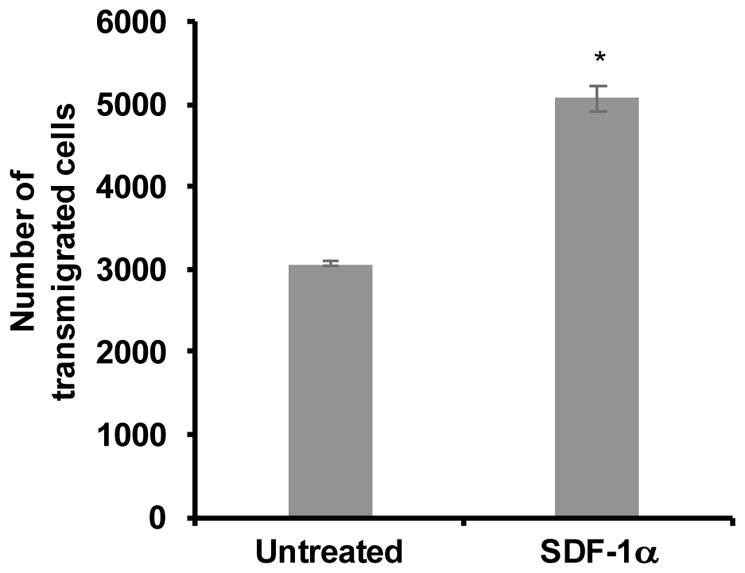
hCMEC/D3 cells were cultured for 10 days on porous PTFE transwell inserts (3 μM). Separately, PBMCs were maintained in non-adherent culture in the presence of IL-2 for 24 hours. At confluence of the monolayer, the CMEC media was removed from both chambers, new co-culture media was added to the lower chambers, in the absence or presence SDF-1α (100 ng/mL), and a suspension of 1 x 10⁶ PBMCs in co-culture media was added to the upper chambers. Transmigration was allowed to occur over a period of 24 hours during incubation at 37°C in 5% CO2. Following transmigration, cells were collected from the lower chambers and analyzed by flow cytometry. Cells were characterized based on forward and side scatter, to identify the PBMC population, and quantified. All bars represent the average two independent experiments performed in triplicate treatments +/− standard deviation. All treatments were significantly increased when compared to untreated as determined by student’s t-test. *p < 0.0013