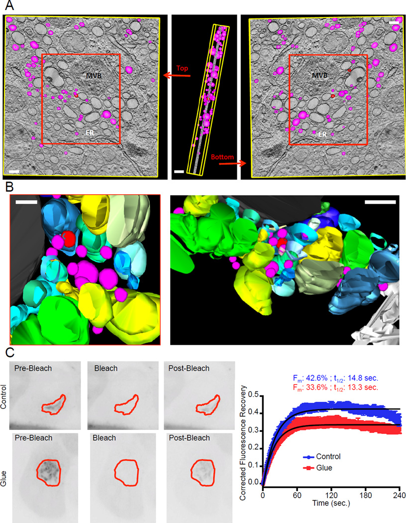
Figure 3. Vesicles form in glued Golgi.
A–B. Electron tomography was performed on HeLa cells expressing DsRed-FRB-Golgin97, Grasp55-FKBP3-CFP and Grasp65-FRB-YFP, treated with Dimerizer for 16 hours. 3 dimensions reconstruction was performed on 81 tilt series using the IMOD software. Golgi cisternae are represented in shades of blue, green and yellow. The colors were used to facilitate the distinction between each reconstituted partly swollen cistern. Vesicles and vesicle buds (modeled as spheres of 50 to 150 nm in diameter entirely enclosed in the thickness of the section) are magenta and red respectively. A multi-vesicular body is colored black and the endoplasmic reticulum (ER) is white. For clarity purposes, only vesicles were modeled in A, while all the structures shown in the red box were modeled in B. Scale bars: 150 nm. C. Fluorescence Recovery After Photobleaching experiments were performed on HeLa cells expressing DsRed-FRB-Golgin97, Grasp55-FKBP, Grasp65-FRB and Arf1-GFP. GFP fluorescence was bleached and its recovery monitored in the Golgi region. Data was single-corrected and averaged over at least 5 cells taken from two independent experiments. Data was fitted on a one-phase association curve (black line).

