
Figure 5. Platelet Adhesion Assay on Rapid-Seeded Sintered Ti that Had a Confluent Cell Coating.
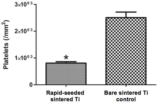
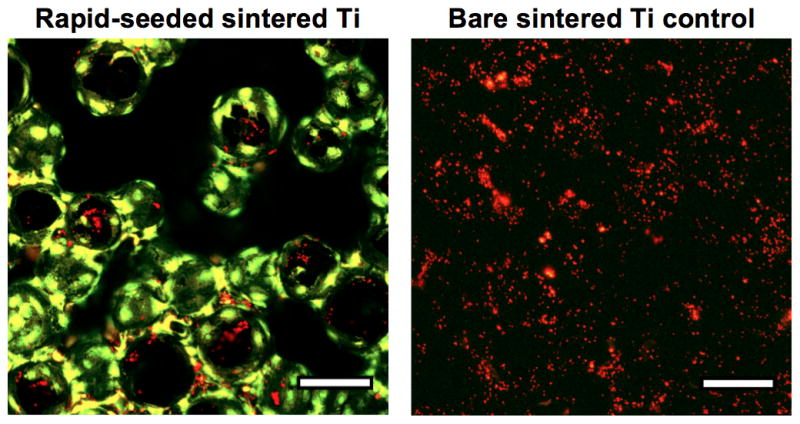
(A) The quantification and (B) representative confocal images of platelets on rapid-seeded sintered Ti after the hCB-ECs had formed a confluent monolayer. (Rapid-seeding density: 0.9 × 10⁵ cells/cm2; n=3; green: ECs; red: platelets; error bars: SEMs; black holes in confocal images were due to different focal plane layers, scale bars: 100 μm.)