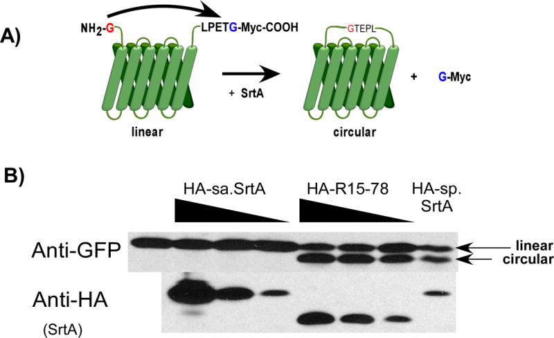
Figure 6.

GFP circularization assay to detect intracellular sortase activity. (A) A GFP construct bearing an N-terminal glycine and C-terminal LPETG peptide is transfected into cells. In the presence of sortase activity, the LPETG peptide is cleaved and attached to the glycine at the N-terminus resulting in a circularized protein. This circularized product can be detected via Western blot as a band of lower apparent molecular weight. (B) HEK293T cells were transfected with the GFP construct along with HA tagged wild type sortase A from S. aureus (HA-sa.SrtA), mutant R15-78 (HA-R15-78) or wild type sortase A from S. pyogenes (HA-sp.SrtA). The production of circularized GFP was assessed by Western using an anti-GFP antibody. Production of HA-sa.SrtA, HA-R15-78 or HA-sp.SrtA was detected using anti-HA antibody. The linear and circular GFP products are indicated.
