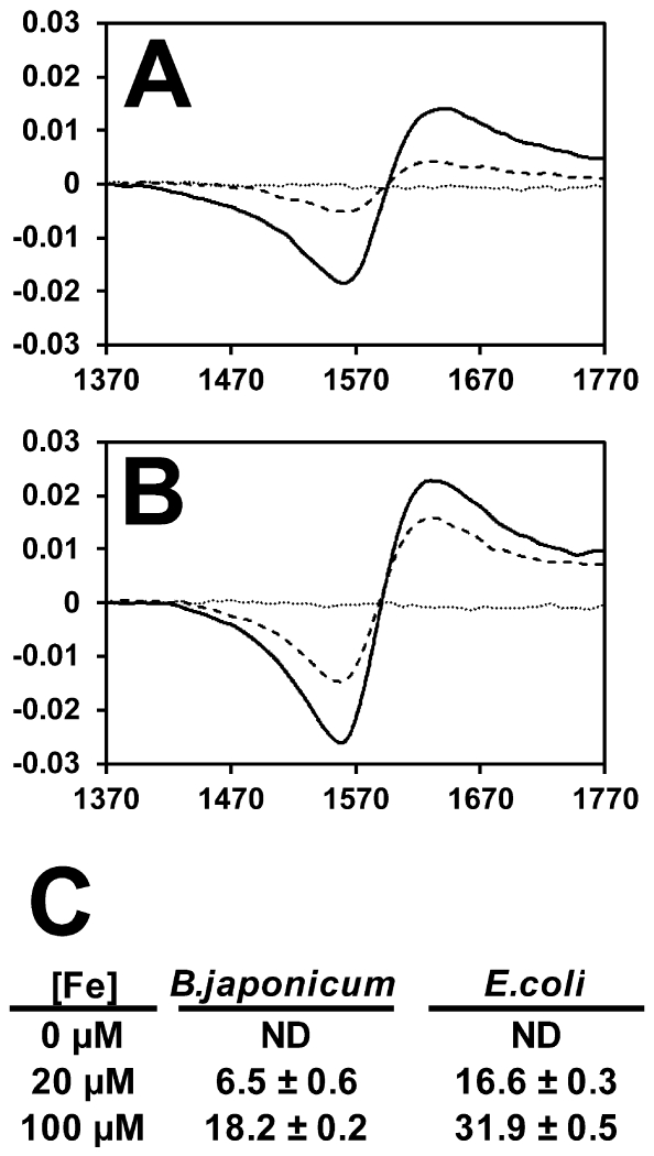
Fig 7.
Determination of intracellular chelatable iron in B. japonicum and E. coli cells by EPR.
EPR raw data traces for (A) B. japonicum or (B) E. coli cells treated with the iron chelator desferrioxamine after growth in media containing no added iron (dotted line), 20 μM FeCl3 (dashed line) or 100 μM FeCl3. (solid line) FeCl3. (C) Quantitation of intracellular chelatable iron as determined using EPR normalized to protein content. Data are expressed as average and standard deviation of triplicate trials. The data are expressed as nmol Fe per mg protein.

