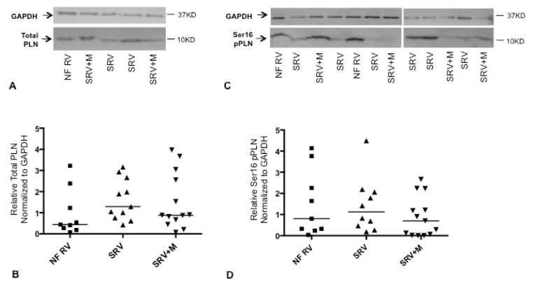
Figure 5.
Total phospholamban (A and B) and phospholamban phosphorylation at the serine 16 residue (C and D) in nonfailing pediatric right ventricle and SRV; representative Western blot and quantitation for each are shown. Horizontal lines in B and D represent population medians; scatter plot and median shown due to non-normally distributed data. GAPDH was used as a loading control. Ser16, serine 16 residue; pPLN, phospholamban phosphorylation; PLN, phospholamban; GAPDH, glyceraldehyde 3-phosphate dehydrogenase; NF, nonfailing (n=9); SRV, single right ventricle (n=11); SRV+M, single right ventricle treated with milrinone (n=13).

