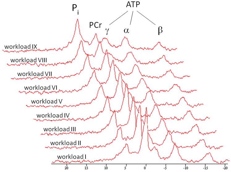
Fig. 1.

Typical time series of 31P NMR spectra acquired from the medial head of the quadriceps muscle of the right leg of a study participant performing incremental exercise to exhaustion. Each spectrum corresponds to three summed free induction decays collected during the final 36 s of each 1 minute workload. A 10 Hz line broadening filter was applied prior to Fourier transformation. Pi = inorganic phosphate; PCr = phosphocreatine; ATP = adenosine triphosphate.
