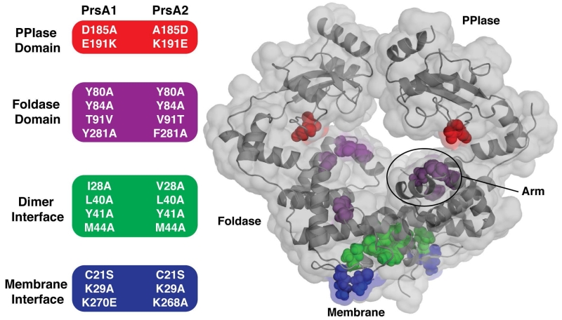
Figure 4. Summary of PrsA1 and PrsA2 variants tested in vivo.
(Left) The residues selected for substitution in both PrsA1 and PrsA2 are grouped and color-coded by domain and/or structural feature. (Right) Transparent surface representation of PrsA1 with selected variant residues drawn as color-coded spheres. PPIase domain substitutions (red), Foldase domain substitutions (purple), dimer interface substitutions (green), and membrane interface substitutions (blue).

