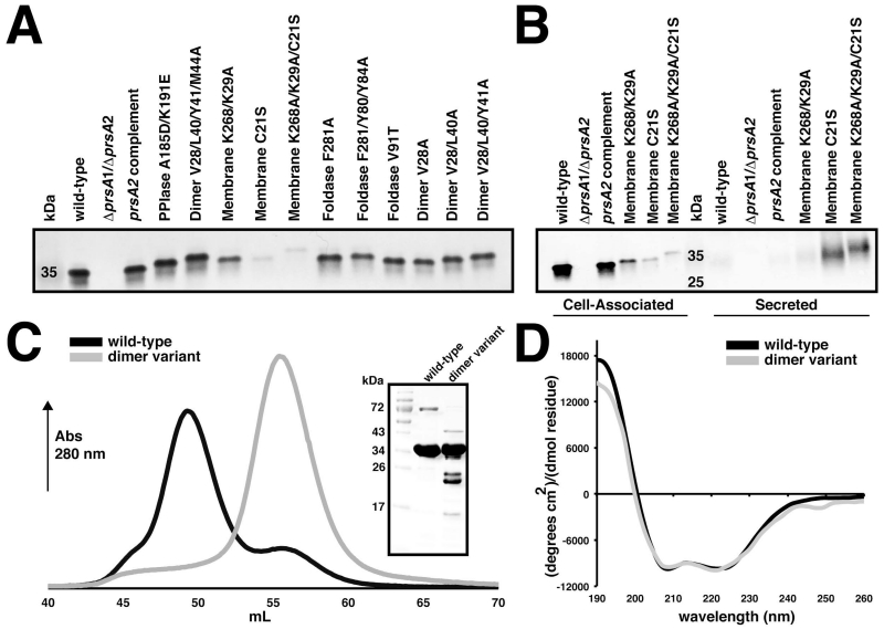
Figure 5. Mutant strain and protein variant control assays.
(A) Cell associated protein profiles for PrsA2 variants expressed in the ΔprsA2/ΔprsA1 background. (B) Secretion profiles of PrsA2 membrane variants in the ΔprsA2/ΔprsA1 background. Bacterial pellets were collected at mid-log phase to detect cell associated protein expression, whereas secreted proteins were precipitated from the media. In both cases PrsA2 variants were visualized by western blot with anti-PrsA2 antibody. (C) Gel-filtration profile of wild-type PrsA1 (black) and the PrsA1 dimer variant (V28/L40/Y41/M44A). Dimerized PrsA1 elutes at ~ 49 mL and monomeric PrsA1 at ~ 55 mL. The inset shows purified wild-type and variant PrsA1 separated by SDS-PAGE gel visualized by Coomassie stain. (D) Circular dichroism spectra of PrsA1 wild-type and the dimer variant plotted as mean residue ellipticity.

