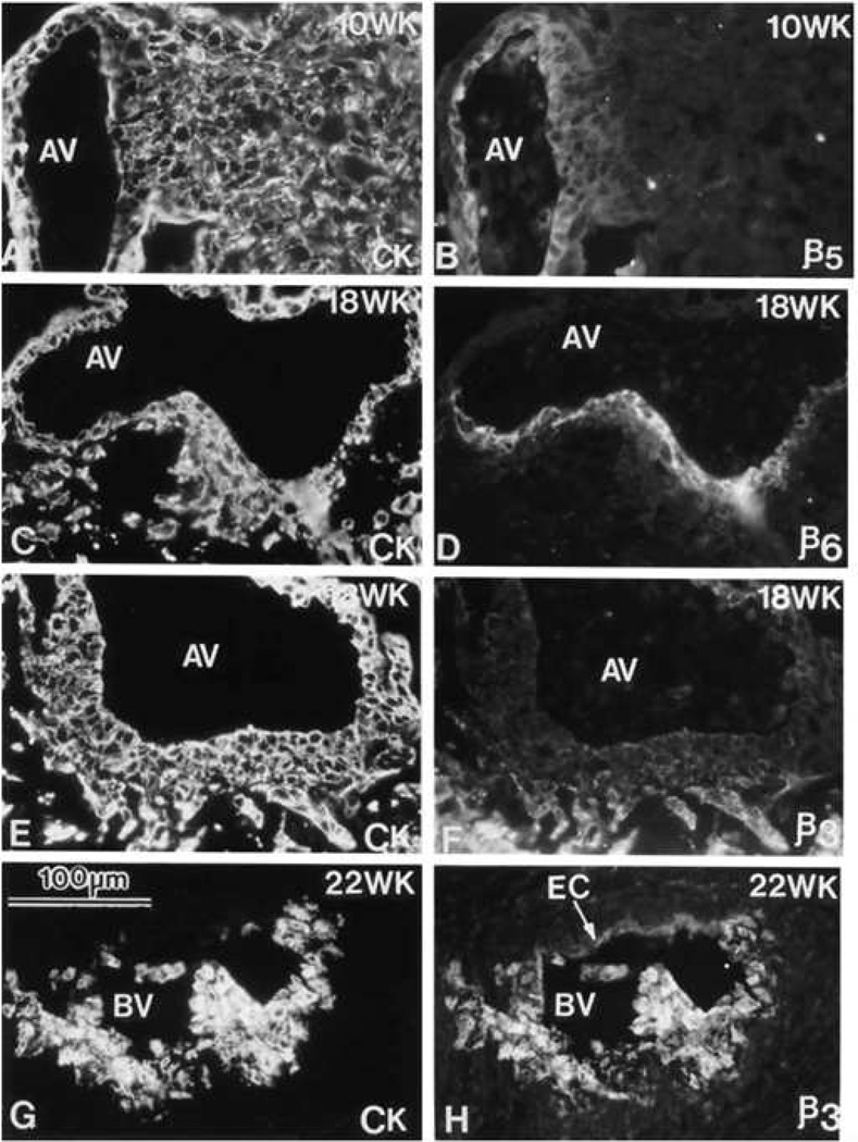
Figure 2. Phenotypic transformation of cytotrophoblast during uterine invasion.
Cytotrophoblasts (CTBs) switch their expression of integrin αVβ (αVβ) family members as they invade the uterine wall. Sections of the maternal-fetal interface at various weeks (WK) of gestation (18–22) were double stained with anti-cytokeratin (CK) to mark CTBs (panels A, C, E and G) and anti-αVβ5 (β5), anti-αVβ (β6), or anti-αVβ3 (β3) (panels B, D, F, and H, respectively). αVβ5 was detected on CTBs in floating (data not shown) and anchoring villi (AV), but not in other locations. αVβ6 was detected on villous CTBs at sites of column formation and in the first cell layer of the column. αVβ3 was upregulated in the distal portions of the columns and on endovascular CTBs that lined the maternal blood vessels (BV). EC, endothelial cell. Reprinted, with permission, from Zhou et al.134

