
Fig. 5.
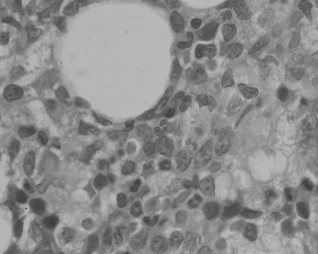
Hematoxylin and eosin-stained sections of trephine biopsy section from patient 2 showing sinusoidally arranged large cells (×400 magnification)
Official websites use .gov
A
.gov website belongs to an official
government organization in the United States.
Secure .gov websites use HTTPS
A lock (
) or https:// means you've safely
connected to the .gov website. Share sensitive
information only on official, secure websites.
Hematoxylin and eosin-stained sections of trephine biopsy section from patient 2 showing sinusoidally arranged large cells (×400 magnification)