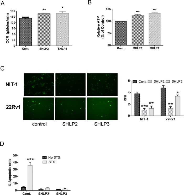
Figure 3. SHLP2 and SHLP3 modulate mitochondrial function.
The effects of exogenous SHLP2 and SHLP3 on mitochondria were assessed in 22Rv1 cells incubated with 100 nM control peptide, SHLP2, or SHLP3 for 24 h by measuring (A) oxygen consumption rate (OCR) performed on a Seahorse XF24 Extracellular Flux Analyzer and (B) ATP production. (C) Reactive oxygen species (ROS) production as assessed by DHE (dihydroethidium) fluorescence in NIT-1 (top) and 22RV1 (bottom) cells after incubation with 100 nM control peptide, SHLP2, or SHLP3 overnight. All data are presented as means ± SEM. (D) NIT-1 β cells were pre-incubated with 100 nM SHLP2 or SHLP3 for 5 h, followed by incubation with 10 μM staurosporine (STS) for 24 h. Apoptosis (pre-G1 peak) was assessed by FACS (fluorescence-activated cell sorting) analysis. *P < 0.05; **P < 0.01; ***P < 0.001.

