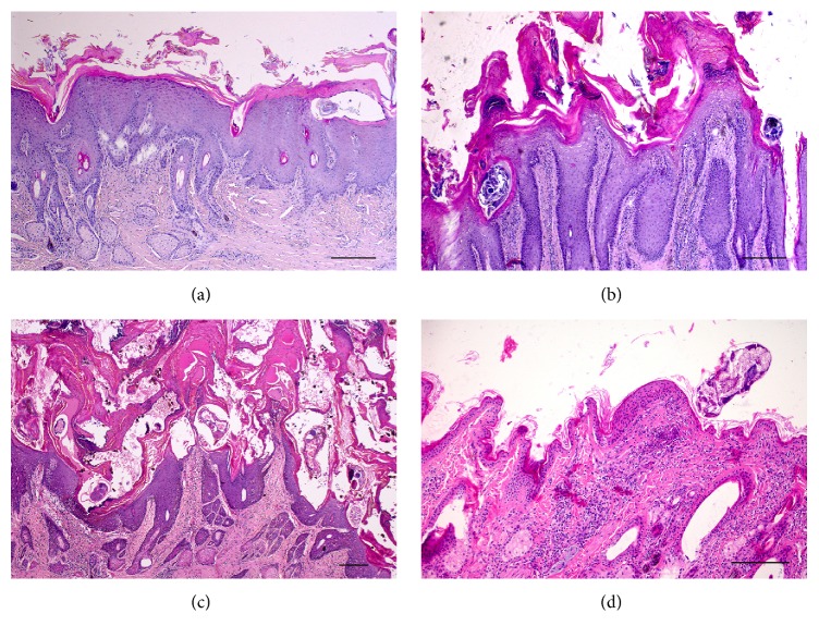
Figure 1.
Histology of skin in sarcoptic (a: grade 1; b: grade 2; c: grade 3) and trombiculosis (d) affected chamois. Severe epidermal hyperplasia is evident in all grades of sarcoptic mange lesions with females within epidermal layers. Crusting with marked parakeratosis is progressively severe from grade 1 to grade 3. In grade 3 (c), crusts are associated with serum lakes, extravasated erythrocytes, and bacteria. Diffuse inflammatory infiltrates are evident in the dermis. Focal, mild epidermal hyperplasia is evident in trombiculosis (d) with a mite on the surface of the keratin layer and no crusts. Focal inflammatory infiltrates are evident in the dermis. Haematoxylin Eosin; bar = 100 μm.

