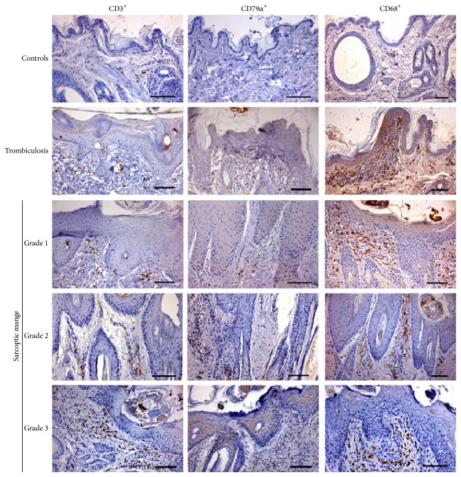
Figure 2.
Immunohistochemical staining of skin of control, trombiculosis, and mange affected chamois. Scanty T lymphocytes (CD3+ cells) and macrophages (CD68+ cells) and very rare B lymphocytes (CD79α + cells) are evident in control chamois. Focal inflammatory infiltration with a prevalence of macrophages is evident in trombiculosis-affected chamois. A prevalence of macrophages and T lymphocytes is evident in all three groups of sarcoptic mange affected chamois. DAB chromogen and haematoxylin counterstain. Bar = 100 μm.

