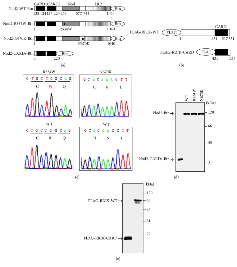
Figure 1.
Schematic representation of Nod2 and RICK, and sequencing chromatograms of Nod2 plasmids containing the BS/EOS-associated mutations, and protein syntheses. (a) Schematic representations of biotinylated wild-type and mutant Nod2. C-terminal biotinylated full-length wild-type Nod2 (Nod2-WT-Btn), full-length R334W-mutated Nod2 (Nod2-R334W-Btn), full-length N670K-mutated Nod2 (Nod2-N670K-Btn), and tandem CARD1- and CARD2-domains-only Nod2 (Nod2-CARDs) are indicated. (b) Schematic representations of FLAG-tagged wild-type and CARD-domain-only RICK. N-terminal FLAG-tagged full-length RICK (FLAG-RICK-FL) and CARD-domain-only RICK (FLAG-RICK-CARD) are indicated. The caspase recruitment domain (CARD) is indicated by black boxes. The nucleotide-binding oligomerization domain-containing protein (Nod) is indicated by grey boxes. Leucine-rich repeats are indicated by striped boxes. Amino acid sequence number and mutated amino acids are indicated under each schema. (c) Sequencing chromatograms of Nod2 and mutated-Nod2 plasmids. The wild-type and mutated-Nod2 plasmids pDONR221-Nod2-WT, pDONR221-Nod2-R334W, and pDONR221-Nod2-N670K were sequenced to confirm (from CGG to TGG corresponding to R334W in the right panel; from AAC to AAA corresponding to N670K in the left panel) mutations at the appropriate site. (d) Western blotting analysis of biotinylated Nod2 and its mutants. A total of 1.5 μg of synthetic protein was subjected to SDS-PAGE followed by Western blotting. Protein detection on the membranes was performed using HRP-conjugated streptavidin. Molecular weights are indicated at right. (e) Western blotting analysis of FLAG-tagged RICK and CARD-domain-only RICK. A total of 1.5 μg of synthetic protein was subjected to SDS-PAGE followed by Western blotting. Protein detection on the membranes was performed using anti-FLAG mAb M2. Molecular weights are indicated at right.

