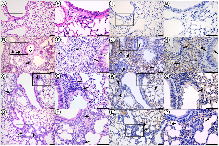
Figure 5. Histopathoglogical and immunohistochemical finding in the lungs of infected mice at 5 d.p.i.
Lung histopathology sections (magnification, 200×) of mice were shown at 5 d.p.i. for the (A) PBS mock-infection control, (B) wild type, (C) CypA-SPC, and (D) CypA-CMV groups. (E–H) Enlargements (600×) for panels (A–D), respectively. Inflammatory cell infiltration, deciduous epithelium mucosae and inflammatory cells in the bronchial lumen, as well as hemorrhage are denoted with thick black arrows, thick white arrows, and black triangles, respectively. (I–L) Immunohistochemically stained sections (200×) corresponding to the histopathology sections, respectively. (M–P) Enlargements (600×) for panels I to L, respectively. Positive signals are denoted with thick solid arrows. Scale bar = 100 μm.

