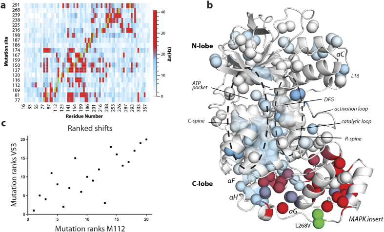
Figure 1. Long-range methyl chemical shift perturbations.
(a) Heat-map of 1H-13C chemical shift perturbations (in Hz) for methyl resonances in p38γ (mutation-wildtype) caused by minimally disruptive mutations. Green represents mutation site. (b) 1H-13C chemical shift perturbations due to the substitution L268V in the MAPK insert mapped onto a homology model of the inactive apo p38γ structure. Colors correspond to shift differences in Hz, using the same scale in (a). (c) The ranks of each mutation induced chemical shift perturbation for V53 and M112. Each point corresponds to a mutant as ranked among the set of mutations for the specified residue.

