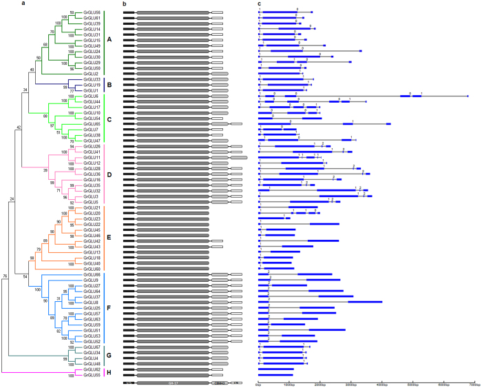
Figure 3. Phylogenetic relationships, protein domain architecture and gene structures of β-1,3-glucanases in cotton.
(a) The multiple alignment of the conserved glycosyl hydrolase family 17 domain were constructed with Clustal X (version 2.0)49, and gaps and poorly aligned sections were removed (Supplementary Fig. 3). Phylogenetic tree was generated using the maximum likelihood method under WAG model in MEGA v5.250, and the reliability of interior branches was assessed with 1000 bootstrap resamplings. (b) Protein domain architectures as defined in Fig. 2. (c) The gene structures were drawn using the online tool GSDS. Introns and exons were represented by black lines and blue boxes, respectively, and numbers at the exon-intron joints were intron phases.

