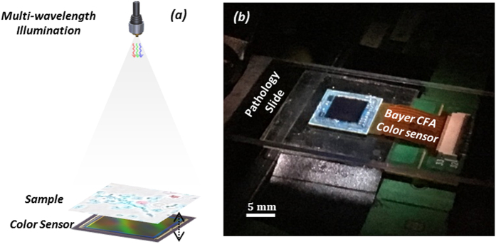
Figure 1. Wavelength-multiplexed holographic color imaging setup.
(a) Schematics of the set-up. The sample (e.g., a pathology slide) is placed ~6 cm below the illumination fiber aperture and its in-line transmission hologram is captured by a Bayer color sensor chip that is placed at <1 mm below the sample. The sensor is mounted on a mechanical stage for 3D motion. (b) Photo of the same on-chip holographic imaging set-up.

