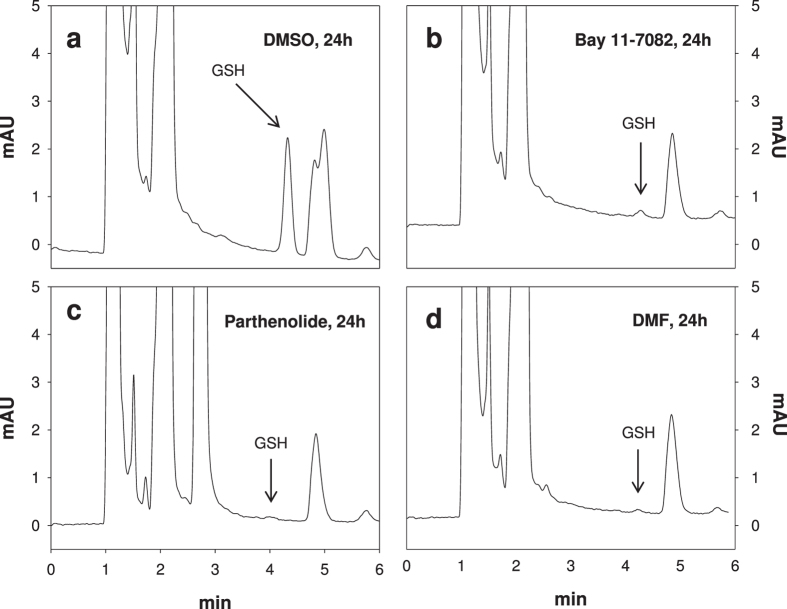
Figure 2. Representative chromatograms obtained for the analysis of GSH in erythrocytes.
The GSH-NEM conjugate was analyzed by reverse-phase HPLC with ultraviolet detection at 265 nm (r.t. 4.36 min). GSH depletion in erythrocytes treated with DMSO (0.2% v/v), Bay 11–7082 (20 μM), parthenolide (50 μM) or dimethyl fumarate (DMF: 140 μM) for 24 h are shown.

