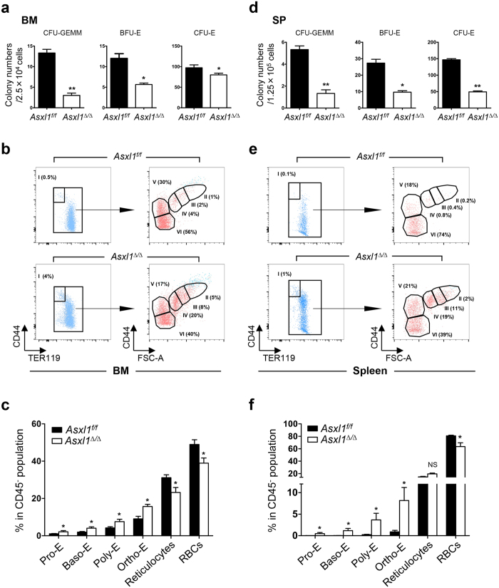
Figure 3. Loss of Asxl1 impairs erythroid terminal maturation.
(a,d) Colony forming unit (CFU) assays with BM cells and spleen (SP) cells from Asxl1∆/∆ and Asxl1f/f mice (3 mice/genotype from two independent experiments). (b,e) Representative flow cytometry dot plots show the percentage of various erythroid subsets in the BM (b) and SP (e) of Asxl1f/f and Asxl1∆/∆ mice. Dot plots in (b,e) show CD45−Mac1−Gr.1− gated cells. Pro-E (CD44hiTER119low, population I), Baso-E (population II), Poly-E (population III), Ortho-E (population IV), reticulocytes (population V), and red blood cells (population VI) groups were identified according to their forward scatter (FSC) and CD44 expression. (c,f) Quantitation of the erythroid populations in the BM (c) and SP (f) of Asxl1f/f and Asxl1∆/∆ mice (n = 5 mice/genotype). Data were presented as mean ± SEM. *p < 0.05, **p < 0.01.

