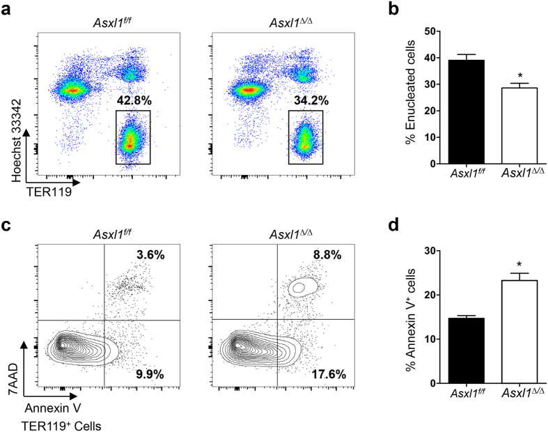
Figure 4. Loss of Asxl1 impairs enucleation and increases the apoptosis of erythroid cells in vivo.
BM cells from Asxl1f/f and Asxl1∆/∆ mice were stained with TER119/Hoechst 33342 or 7AAD/Annexin V and flow cytometric analysis was performed to quantify erythroid enucleation or apoptosis. (a) Representative dot plots show the percentage of erythroid enucleation in the BM of Asxl1f/f and Asxl1∆/∆ mice. (b) Quantification of enucleated erythroid cells shown in (a) (TER119+ and Hoechst 33342−, n = 5 mice/genotype). (c) Representative contour plots of apoptosis show early and late apoptotic cell populations (7AAD−Annexin V+ and 7AAD+ Annexin V+). (d) Quantification of Annexin V-positive cells shown in (c) (n = 3 mice/genotype). Data were presented as mean ± SEM. *p < 0.05.

