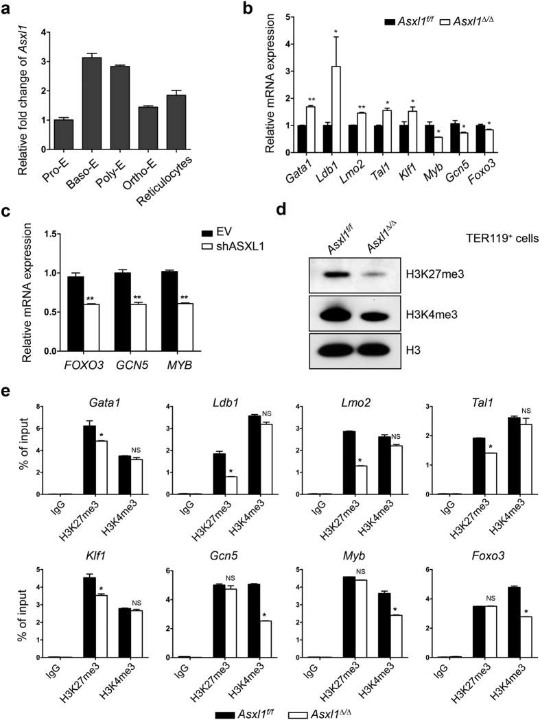
Figure 5. Deletion of Asxl1 dysregulates genes critical for erythropoiesis through altered H3K27me3 and H3K4me3 occupancy in TER119+ cells.
(a) Relative expression level of Asxl1 in purified erythroblast populations. (b) qPCR analysis of critical genes associated with erythroid differentiation and maturation in TER119+ cells from Asxl1∆/∆ and Asxl1f/f (n = 3 mice/genotype). Data are shown as expression units relative to the respective gene expression in Asxl1f/f mice using β-actin as an internal calibrator. (c) The mRNA expression of FOXO3, GCN5, and MYB in the erythroid progenitors of CB CD34+ cells transduced with shASXL1 after 12 days of culture in vitro. Data are shown as expression units relative to the respective gene expression in control cells, using 18S as an internal calibrator. (d) Western blot analysis of H3K27me3 and H3K4me3 in the TER119+ cells from Asxl1∆/∆ and Asxl1f/f mice. The total H3 levels served as a loading control. Representative blots from 3 independent experiments are shown. (e) ChIP-qPCR for H3K27me3 and H3K4me3 at/around the Gata1, Ldb1, Lmo2, Tal1, Klf1, Myb, Gcn5, and Foxo3 TSS regions (n = 3 mice/genotype from two independent experiments). Data are represented as mean ± SEM. *p < 0.05, **p < 0.01.

