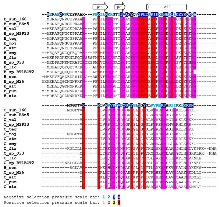
Figure 3.
Multiple sequence alignment of the SpoIISB and SpoIISC amino acid sequences from the B. subtilis group. Residues that are identical within either the SpoIISB or SpoIISC proteins are red, and similar ones are purple. Selection pressure is indicated on the SpoIISB and SpoIISC reference sequences (from B. subtilis strain 168) shown above each sequence block. Selection pressure intensity is indicated by the given scale bar. SpoIISB secondary structure elements (as identified in [7]) are shown above the alignment; the abbreviations are from Table S3.

