Figure 1.
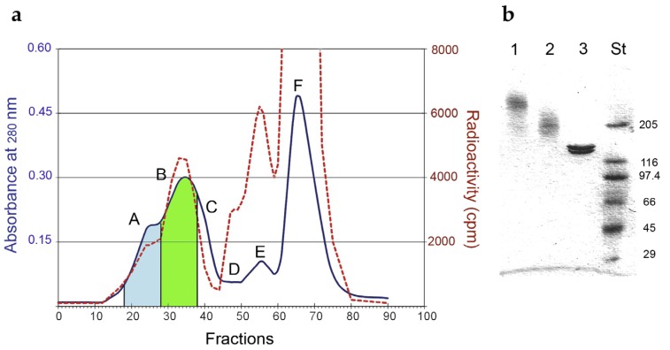
(a) Elution profile of gel-filtration chromatography on a Sephacryl S-200 HR column of Rituximab/saporin-S6 conjugate. The blue line represents the A280, and the red dashed line represents the radioactivity of the eluted fractions. The fractions corresponding to the peaks were pooled and indicated with capital letters: (A) high-molecular-weight immunotoxin (HMW-IT) (blue area); (B) low-molecular-weight immunotoxin (LMW-IT) (green area); (C) free Rituximab; (D) trimmers; (E) dimers and (F) monomers of saporin-S6. (b) Analysis of fractions corresponding to HMW-IT (1), LMW-IT (2), and unconjugated Rituximab (3) by SDS–PAGE under non-reducing conditions on a 4%–15% PhastGel. Standard molecular weights (St) are expressed in kDa.

