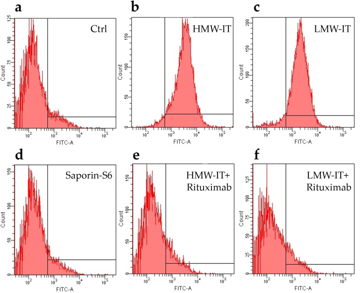
Figure 4.
Immunospecificity of the immunotoxins towards CD20 expressing cells (Raji) by cytofluorometric analysis. The cells were incubated with HMW-IT and LMW-IT in the absence (b,c) or in the presence (e,f) of 100-fold molar excess of Rituximab. The immunotoxins binding was detected by rabbit antisera against saporin-S6, followed by an anti-rabbit FITC antibody. Control samples were run with complete medium alone (see Matherials and Methods) (a) or with saporin-S6 (d).

