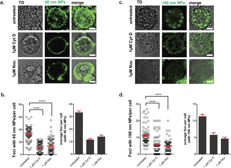
Figure 3. Effect of cytoskeletal inhibitors on uptake of nanoparticles.
(a) Fluorescent confocal 2D micrographs of fresh macrogametocytes incubated in the presence of fluorescent 40 nm nanoparticles prior to and after the addition of cytoskeletal inhibitors, cytochalsin D and nocodaloze. Intracellular 40 nm NPs are shown in green. Scale bar: 5 μm. (b) Quantification of foci containing intracellular 40 nm particles in cells with and without cytochalasin D and nocodazole (mean ± range, n = 3, P = two-tailed t-test). (c) Confocal micrographs showing uptake of 100 nm particles (particles: green) in untreated and drug-treated macrogametocytes. Scale bar: 5 μm. (d) Quantification of foci containing intracellular 100 nm particles in cells with and without cytochalasin D and nocodazole. Mean ± range, n = 3, P = two-tailed t-test.

