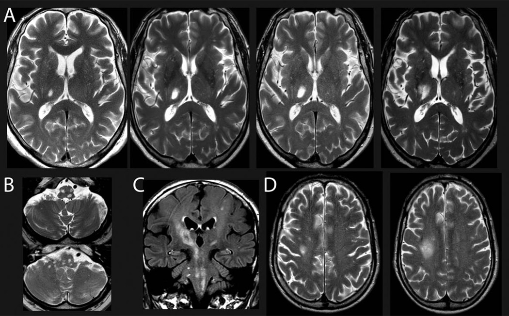
FIGURE 1.
(A, B) Representative magnetic resonance imaging showing the progressive enlargement of the lesion in the right thalamus (A; March 2013, July 2013, December 2013, March 2014) and multiple lesions in the mesencephalon, pons, cerebellar pedunculi, and medulla oblongata (B; March 2014) within the first 1.5 years after onset of neurological symptoms by T2-weighted imaging. (C) Fluid-attenuated inversion recovery imaging showing widespread and diffuse affection of the midbrain structures including the pyramidal tract and pontine and cerebellar structures (March 2014). (D) T2-weighted imaging (September 2014, December 2014) shows a progressive subcortical lesion that was chosen as biopsy target (see Fig 3). For time points of magnetic resonance imaging, please see also Figure 2.

