Figure 2.
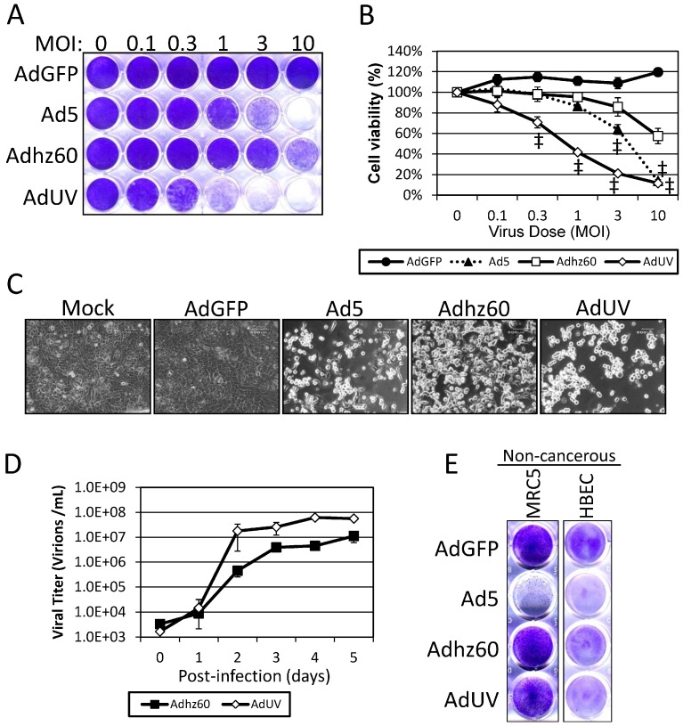
AdUV is released from and lyses A549 cells more efficiently than Adhz60. (A) A549 cells stained with crystal violet after five days’ treatment with the indicated Ads and MOIs; (B) Quantification of A549 cells stained with crystal violet expressed at the percent (%) viability; (C) Cytopathic effects (CPE) in A549 cells treated at an MOI of 1 for the indicated Ads were photographed at 200× total magnification five days post-infection; (D) A549 cells were infected with at an MOI of one with Adhz60 and AdUV and media samples were collected daily until day 5. Ad titer was determined via the TCID50 method using HEK293 cells. Day zero represents samples collected at 6 h post-infection; (E) Crystal violet staining of non-cancerous lung fibroblasts (MRC5) and epithelial (HBEC) cell lines treated at an MOI of 10 for three days. Quantified data are shown ± the standard deviation (SD) of three independent replicates. Statistical significance was assessed via two-way ANOVA relative to Adhz60 treated cells with multiple comparisons corrected for by Bonferroni’s method. ‡ indicates p-value > 0.001.

