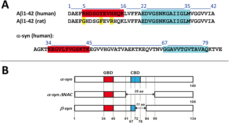
Figure 1. Amino acid sequence and lipid-binding domains in Aβ, α-synuclein and related proteins.
(A) Amino acid sequence of Aβ1-42 (human and rat) and of the region of human α-synuclein that contains the ganglioside-binding domain (in red) and the cholesterol-binding domain (in blue). Non-conserved amino acid residues in rat Aβ1-42 are in yellow. Note that both the cholesterol-biding domain (fragment 67–79) and the tilted peptide of α-synuclein (67–78) bind cholesterol with high affinity. (B) Alignment of α-, β-, and ΔNAC synucleins. Note that all three proteins share the same ganglioside-binding domain (GBD, in red) whereas only α-synuclein displays a complete cholesterol-binding domain (CBD, in blue).

