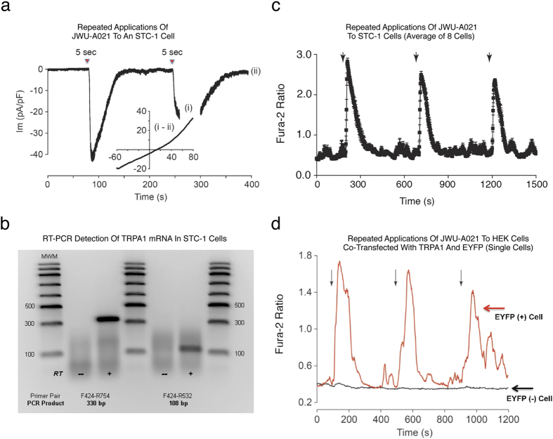
Figure 6. Membrane currents and Ca2+ transients activated by JWU-A021.
(a) Whole-cell patch clamp analysis (Vh −60 mV) demonstrated inward membrane currents activated by repeated 5 sec focal applications of JWU-A021 (3 μM; red triangles) to a single STC-1 cell. The inset provides a current-voltage (I-V) relationship for the current activated by JWU-A021 (Im, membrane current in pA normalized to membrane capacitance in pF). It is the difference current obtained by subtracting the IV relationships measured during (time point “i”) and after recovery (time point “ii”) of the response. Findings are representative of a single patch clamp experiment that was repeated with similar results using N = 10 cells. (b) RT-PCR validation that STC-1 cells express TRPA1 channel mRNA, as detected using two different primer pairs (RT, reverse transcriptase; MWM, molecular weight markers). Findings are representative of a single experiment repeated twice with similar results. (c) Averaged Ca2+ transients obtained from STC-1 cells stimulated by focal application (arrows) of JWU-A021 (3 μM) to N = 8 cells. (d) Ca2+ transients stimulated by focal application (arrows) of JWU-A021 (3 μM) to a single HEK-293 cell transfected with rat TRPA1 cDNA fused to EYFP cDNA (red trace), or a HEK-293 cell transfected with EYFP cDNA but not rat TRPA1 cDNA (black trace). EYFP fluorescence was used as a marker to positively identify cells that were transfected so that fura-2 based assays of [Ca2+]i could be performed using these cells. Findings are representative of a single experiment repeated a minimum of three times on three different occasions with similar results.

