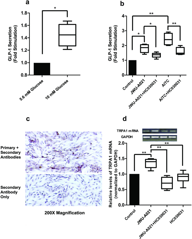
Figure 8. JWU-A021 stimulates GLP-1 release from mouse intestinal cells.
(a) Primary cultures were stimulated for 30 min using serum-free DMEM assay buffer containing either 5.6 or 10 mM glucose so that glucose-stimulated GLP-1 secretion could be measured. Data are the mean + s.d. of 4 independent assays (*p < 0.05; paired t test) and are expressed as the fold-stimulation of GLP-1 release, so that a value of 1.0 corresponds to GLP-1 release measured for buffer containing 5.6 mM glucose. (b) HC030031 (10 μM) inhibited the actions of JWU-A021 (3 μM) and AITC (100 μM) to stimulate GLP-1 secretion from primary cultures. HC030031 was administered 15 minutes prior to addition of JWU-A021 or AITC, and it was also present during the 30 minutes test interval during which cells were exposed to JWU-A021 or AITC dissolved in serum free DMEM assay buffer containing 5.6 mM glucose. Data are the mean + s.d. of 4–6 independent assays (*p < 0.05; **p < 0.01; ANOVA with Bonferroni post test). (c) Immunocytochemical detection of GLP-1 in primary cell cultures. The top panel illustrates specific GLP-1 immunoreactivity (brown), as detected using the anti-GLP-1 monoclonal primary antibody in combination with an HRP conjugated secondary antiserum. The bottom panel illustrates negative control non-specific labeling obtained when using the secondary antiserum only. (d) qRT-PCR analysis demonstrated that JWU-A021 (3 μM) increased the relative abundance of TRPA1 channel mRNA in primary cell cultures, and that this effect was reduced by HC030031 (10 μM). For this analysis, cultures were maintained for 30 minutes in serum-free DMEM assay buffer containing 5.6 mM glucose and the test compounds. Data are the mean + s.d. of 6 independent assays (**p < 0.01; ANOVA with Bonferroni post test). The top inset illustrates qRT-PCR products detected by agarose gel electrophoresis.

