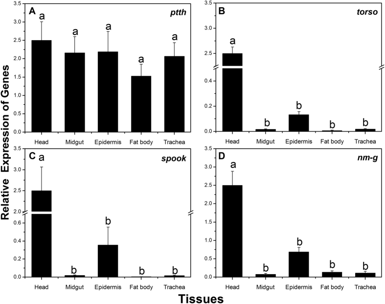
Figure 4. Relative expression levels of ptth (A), torso (B), spook (C), nm-g (D) in different tissues of the 3rd instar larvae of Chilo suppressalis.
Expression levels in the head, midgut, cuticula, fat body, and trachea were detected by q-PCR. Different letter shows significant difference (P < 0.05, ANOVA).

