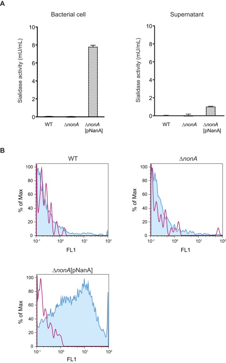
Figure 4. NanA degrades terminal sialic acid displayed on GBS polysaccharide capsule.
(A) Sialidase activities of GBS cells and culture supernatant. After 2 h incubation at 37 °C, fluorescence of sialidase-degraded substrate was measured with excitation and emission wavelengths of 350 and 460 nm, respectively. Data are presented as the mean of sextuplets samples. S.E. values are represented by vertical lines. The sensitivity is 0.3 mU/mL. (B) FITC-labeled ECA binding to live GBS. Red line and blue histogram represents data for bacterial strains incubated without or with ECA, respectively.

