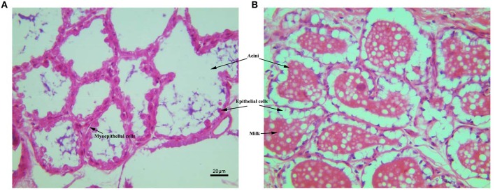
Figure 2.
The histological structure of mammary gland stained with HE. (A) Mammary gland of non-transgenic goat (NG); (B) mammary gland of GH transgenic goat (TG). The Mammary glands of NG and TG were collected in the same lactation period, then fixed and cut into paraffin section. The tissues were stained with HE. The milk, aicni and myoepithelial cells underneath the epithelial cells were labeled.

