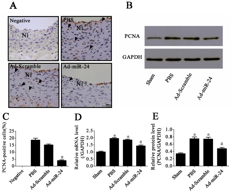
Figure 4.
Anti-proliferative effect of miR-24: (A) Representative microphotographs of immunohistochemistry for proliferating cell nuclear antigen (PCNA) (400× magnification). Arrows indicate positive cells for PCNA. Ni: Neointima. Scale bar represents 20 μm; (B) Representative immunoblots of PCNA; (C) PCNA-positive rate in the neointima (compared to Ad-Scramble group and PBS group, # p < 0.05, n = 6); (D) The mRNA level of PCNA in carotid artery (compared to Sham group, * p < 0.05; compared to PBS group or Ad-Scramble group, # p < 0.05, n = 6); (E) Bar graphs of corresponding densitometric analyses of Western blots (compared to Sham group, * p < 0.05; compared to PBS group or Ad-Scramble group, # p < 0.05, n = 6).

