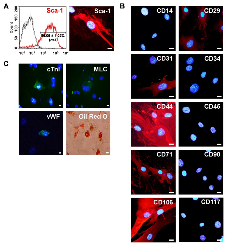
Figure 1.
Isolation of mouse Sca-1+ CSCs from adult heart. (A) Sca-1+ CSCs were enriched by MACS with PE-conjugated anti-Sca-1 antibody and anti-PE micro beads. After sorting four rounds, ~86% of the cells expressed Sca-1 as determined by flow cytometry (left). CSCs expressing intense Sca-1 signals were observed under confocal microscopy after immunostaining with anti-Sca-1 antibodies (right). Scale bars = 20 μm; (B) characterization of Sca-1+ CSCs. Sca-1+ CSCs were stained with anti-CD14, -CD29, -CD31, -CD34, -CD44, -CD45, -CD71, -CD90, -CD106, and CD117 antibodies and visualized with Alexa Fluor 594 secondary antibodies (red). Scale bars = 20 μm; and (C) differentiation potential of Sca-1+ CSCs. Cardiac, endothelial, and adipogenic differentiation were confirmed by immunostaining with cardiomyocyte markers (cTnI, MLC, green), an endothelial marker (vWF, green), and Oil-Red O staining (red), respectively. Nuclei were stained with DAPI (blue). Scale bars = 20 μm.

