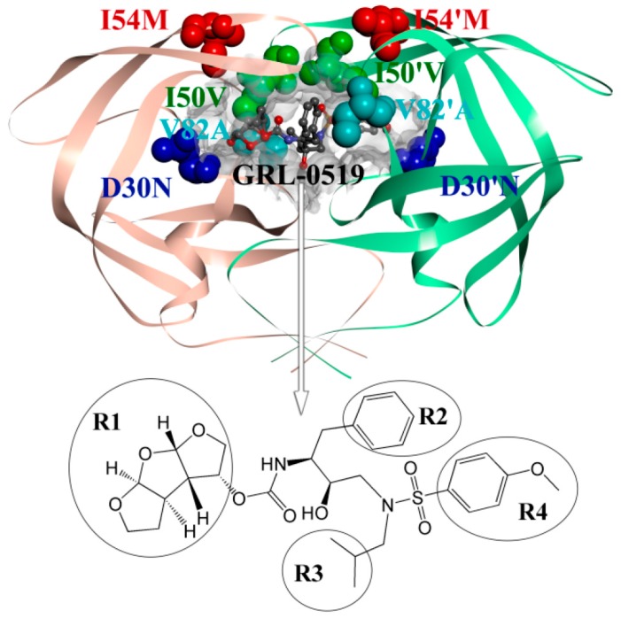
Figure 1.
The locations of the four mutations in the wild type (WT). The PR is displayed in a solid ribbon representation, chain A is shown in light orange, chain B in pale green, GRL-0519 is displayed in a ball and stick representation, and the mutated residues are displayed as a ball with different colors as well as the labeled markers. The molecular structure of HIV-1 PR inhibitor GRL-0519 and the four groups (R1, R2, R3, and R4) are also shown.

