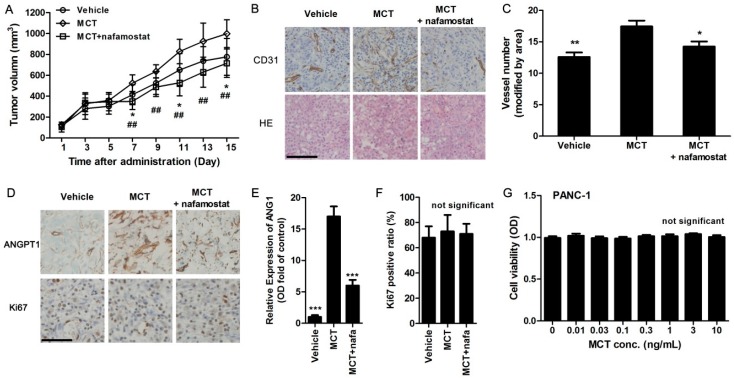
Figure 4.
MCT promotes pancreatic cancer cells tumorigenesis via increasing angiogenesis in vivo. (A) Nude mice bearing pancreatic tumor were subcutaneously injected with MCT (200 ng/kg) or MCT along with nafamostat (10 mg/kg) daily, while vehicle mice received saline. The tumor volume was measured every two days after tumor formed with injection of PANC-1 cells. * p < 0.05 indicated MCT-treated group and vehicle group, ## p < 0.01 indicated MCT-treated group and nafamostat co-treated group, n = 10; (B) After experiments, tumor tissues were removed and embedded for paraffin sections. IHC staining for CD31 and Hematoxylin-Eosin (HE) staining were performed, scale bar = 100 μm and refer to all panels; (C) The microvascular density in tumor tissues was calculated as CD31-positive vessel number (random 10 images, adjusted by tumor areas), * p < 0.05, ** p < 0.01 compared to MCT-treated group; (D) IHC staining for ANGPT1 and Ki67, nuclei were stained with hematoxylin, scale bar = 100 μm and refer to all panels; (E) Optical density (OD) was measured from 10 random images of ANGPT1 staining in each animal, *** p < 0.001 compared to MCT-treated group, n = 10; (F) Ki67-positive ratio was calculated as Ki67-positive nuclei number divided by total hematoxylin-stained nuclei, n = 10; (G) After PANC-1 cells were treated with different concentration of MCT for 24 h, CCK8 assays were used to determine the cell viability, n = 6. All statistical difference were analyzed by ANOVA and Student-Newman-Keuls tests.

