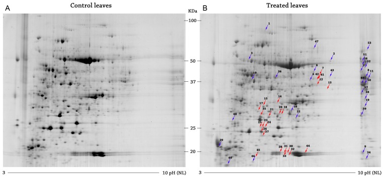
Figure 1.
Representative reference 2-DE gels of Arabidopsis control (A) and treated (B). Gels were colored by colloidal Coomassie blue staining. The Progenesis SameSpot software package was used for gels analysis. The differential proteins are identified by arrows: in red, the down-expressed spots in; in blue, the over-expressed spots in cerato-platanin (CP)-treated leaves. NL, non-linear.

