Figure 1.
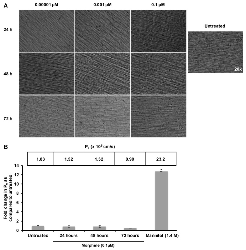
Morphine did not induce leakiness of the hCMEC/D3 barrier. (A) hCMEC/D3 cells were cultured for 10 days on 0.4 µM porous polytetrafluoroethylene (PTFE) transwell inserts. The confluent monolayer was then exposed to morphine (0.1 µM) for 24, 48, or 72 h with morphine additions every 24 h to maintain drug concentration. No changes in cellular morphology were observed. Monolayers incubated in the absence or presence of morphine were visualized under 20× Relief Contrast Microscopy using an Olympus (Center Valley, PA, USA) IX81 deconvolution microscope; (B) hCMEC/D3 cells were cultured and treated as in panel A. Permeability was assessed by determining the amount of 70 kDa fluorescein isothiocyanate–dextran (FITC-D) to pass from the upper to lower chamber over 30 min, and the permeability coefficient (Pe) was calculated. Results show that treatment with morphine does not cause general, non-specific leakiness of the monolayer. All treatments were performed in triplicate and are representative of three independent experiments. Statistical analysis was performed using the analysis of variance (ANOVA) method with log transformation and adjustment for possible time effects to compare the results obtained with untreated monolayers to the results obtained with the morphine-treated monolayers and to the results obtained with monolayers treated with mannitol. Based on 95% confidence intervals, no significant change was observed with morphine treatment. * p < 0.0001.

