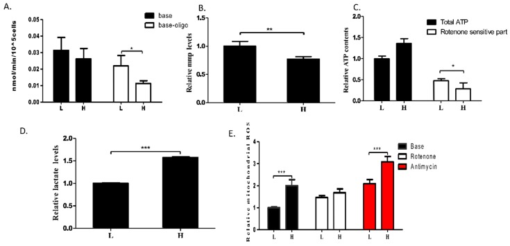
Figure 4.
m.3697G>A mutation-induced OXPHOS defect. (A) Changes in mitochondrial respiratory capacity were determined in clones L and H (n ≥ 3). Oligomycin (2.5 µg/mL) was used to measure OXPHOS-driven mitochondrial respiration, which was calculated by subtracting the uncoupled component from basal endogenous respiration; (B) MMP was determined in clones L and H. MMP levels were measured in cells treated with 30 nM tetramethylrhodamine (TMRM) for 20 min (n ≥ 4); (C) Total ATP content was measured in clones L and H. Rotenone-resistant ATP content was determined in cells treated with 200 nM rotenone for 24 h. Rotenone-sensitive ATP content was calculated by subtracting the rotenone-resistant component from the total ATP content (n ≥ 4); (D) Extracellular lactate levels of clones L and H were measured in the medium after 48 h in culture (n ≥ 4); (E) Mitochondrial ROS levels were determined in clones L and H. Complex I- and complex III-related ROS generation were measured upon inhibition with 1 µM rotenone for 1 h (n ≥ 4) and 5 µM antimycin A for 30 min (n ≥ 4), respectively. The levels of MMP, ATP, lactate, and ROS were normalized against total protein concentration. For (A), 3–4 technical replicates were included; Error bars, ± SD; * p < 0.05; ** p < 0.01; *** p < 0.001.

