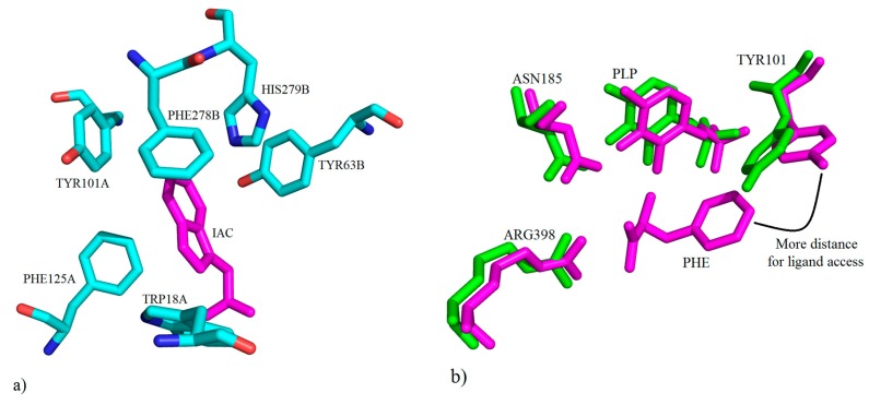
Figure 26.
(a) Presentation of the key molecular interactions between amino acid residues of the active site of hKAT-1 involved in hydrophobic interactions with IAC (shown in magenta) (PDB code: 3FVU); (b) depiction of the native hKAT-1 structure (PDB code: 1W7L) superimposed on the structure of hKAT-1 in complex with PHE (PDB code: 1W7M) to show the conformational change of TYR101’s role.

