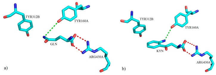
Figure 30.
The three-dimensional representation of key molecular interactions between amino acid residues of the active site of mKAT-3 (a) with GLN (PDB code: 3E2Y) and (b) with KYN (PDB code: 3E2Z). Both ligands form a hydrogen bond with TYR160A via their amino groups (green broken lines). Salt bridge interactions between the ARG430A residue and the carboxylate moiety for both ligands are shown with red broken lines.

