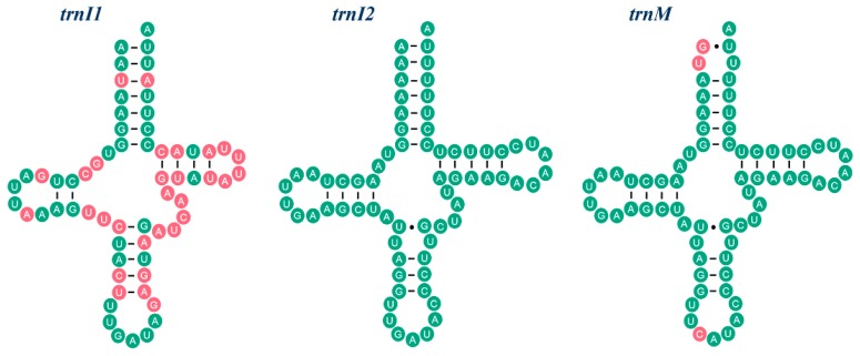
Figure 2.
The secondary stem-loop structure and sequence similarity of trnI1, trnM and trnI2. Inferred Watson-Crick bonds are illustrated by lines, whereas GU bonds are illustrated by dots. Sequences of trnI1 and trnM are compared with trnI2 respectively. The identical nucleotides with trnI2 are labeled with green circles. The variable nucleotides are highlighted in red.

