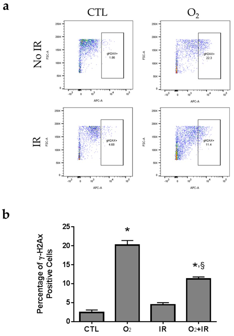
Figure 6.
Flow cytometric analysis of H2AX phosphorylation in lung epithelial cells 48 h (2 cycles) after exposure to hyperoxia, radiation, and double-hit combination challenge. Non-tumorigenic murine alveolar type II epithelial cells (C10) were evaluated after 2 cycles of hyperoxia and radiation exposure and evaluated by flow cytometric analysis of γΗ2ΑΧ and PI. (a) Representative flow cytograms depicting anti-γΗ2ΑΧ (Serine 139) labeling of cells under hyperoxic conditions and IR; (b) Levels of H2AX phosphorylation represented by the percentages (%) of positive stained C10 cells in every cohort. Data are represented as the mean ± SEM. * p < 0.05 for each exposure condition as it compares with the unexposed control (CTL); § p < 0.05 for significant differences between IR-alone compared to O2 + IR.

