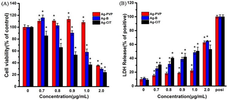
Figure 2.
The viability (A) and LDH level (B) assays of Caco-2 cells after being exposed to three Ag NPs for 24 h. (A) Percent viable cells compared to no particle (0 μg/mL) condition; and (B) percent LDH release compared to 100% cell lysis. All data are represented as the mean ± SD (n = 6). * p < 0.05 comparing with the 0 μg/mL control.

